Introduction
Fungi are eukaryotic, heterotrophic, non-photosynthetic organisms in their own kingdom. The majority is made up of microscopic filaments known as hyphae, and the network of filaments is known as mycelium. They exist as parasites or saprophytes, absorbing organic material from their surroundings. Chitin polymer of the sugar glucosamine is found in their cell walls. Mycelium produces fruiting structures with names like sporangium, ascus, and basidium, to name a few. Sexual or asexual spores can be found in these fruiting structures. The filaments of the hyphae are haploid (1N).
Characteristics of fungi
Also read: Characteristics of fungi
Fungi have distinct microscopic and macroscopic characteristics that distinguish them from other organisms. Most multicellular fungal bodies, also known as molds, are composed of filaments known as hyphae. Hyphae can form a tangled network known as a mycelium, which serves as the thallus (body) of fleshy fungi. Septate hyphae are those that have walls between their cells; non-septate or coenocytic hyphae are those that do not have walls or cell membranes between their cells.
Background
Throughout the 1800s and early 1900s, fungal classification relied heavily on the macroscopic features of the fruiting body, though in the late 1800s, more emphasis was placed on microscopic features. Because microscopic features became more important in the 1900s, while all of the features listed below are still very useful for identification, their role in classification is frequently greatly reduced. The type of fruiting body is extremely important (e.g. mushroom, puffball, cup fungus, polypore, etc)
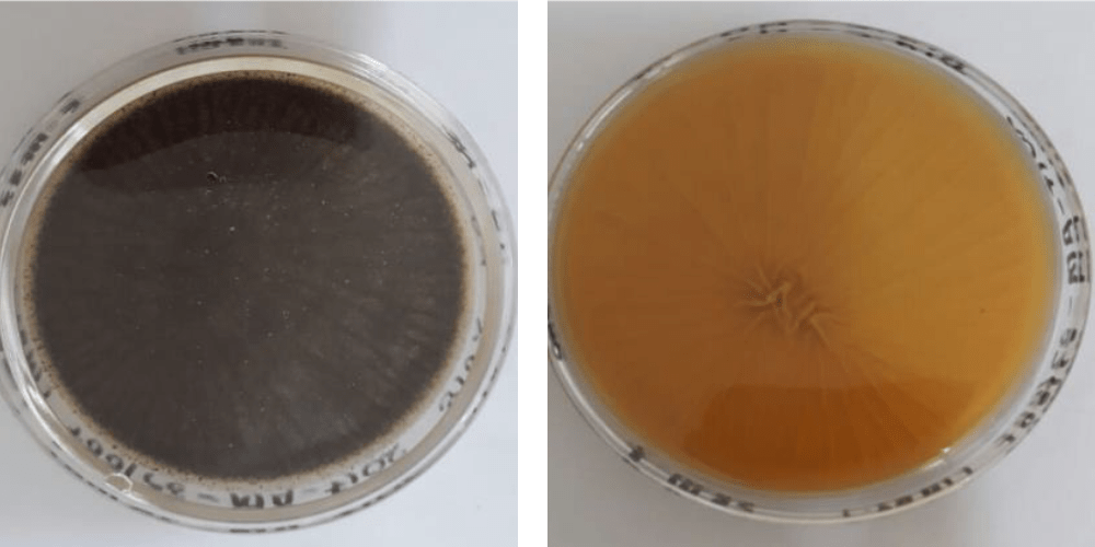
Macroscopic identification: mature and immature colonies of fungi
In macroscopic analysis, colony characters are observed.
- Characters of colony that are observed from the front side of the petri dish are called obverse characters.
- Colony characters observed from the back side of the petri dish are called reverse characters.
These characters are observed on the basis of:
- Surface topography: some fungal colonies grow freely, covering the entire surface of the agar; others grow in a restricted manner.
- Surface texture: fungi can be cottony or woolly (floccose), granular, chalky, velvety, powdery, silky, glabrous (smooth, creamy), or waxy.
- Color: Fungi can be colourless or brightly coloured. Color can be found on the fungus, its sporulating apparatus, the agar, or the colony’s bottom (reverse pigmentation).
- Colony morphology: fungal colonies can be appeared as Rugose, Umbonate, Verrucose, Flat. Texture of fungal colonies can be Cottony, Glabrous, Granular, Velvety
Microscopic characteristics of fungi
If a possible fungus is found in a culture plate, colony characteristics are first evaluated to determine the isolate’s broad group. Following the initial observations, the following microscopic criteria are used to identify the fungal isolate’s genus/species.
Determine the structure of any hyphae
They are as follows:
- Whether septate or aseptate
- Whether to branch (and at what angles) or not to branch
- Pigmented or not
- The width can be even or uneven.
- Arthroconidia or pseudohyphae
- Determine the origin and structure of fruiting bodies.
Consider the following conidiation:
- The spores’ or conidia’s size and shape
- The spores’ size, shape, and arrangement
- Look for the presence of any of the following special diagnostic structures: Hulle cells, pycnidia, and cleistothecia
If you only observe yeast cells:
- take note of their size, shape, and arrangement.
- Examine for the presence or absence of capsules.
- Determine whether the bastoconidiation is single or multiple daughter cells.
Here are microscopic characters that help to identify specific fungi:
- Hyphae are small (3-6 micrometer) and regular in size, branching dichotomously at 45-degree angles with distinct cross-septa: Aspergillus niger
- Hyphae are irregular in size (6-50 micrometer), ribbonlike, and septa-free: Rhizopus-Mucor-Absidia; Zygomycetes (Phycomycetes);
- Hyphae are small (2-3 micrometers) and regular, with some branching and occasional rectangular arthrospores: only found in skin, nail scrapes, and hair: Dermatophyte class (Microsporum specie, Trichophyton specie, Epidermophyton specie)
- Phaeohyphomyces spp., and Hyalohyphomyces spp. have regular diameter hyphae (3-6 micrometers), parallel walls, irregular branching, septate, dark yellow, brown, or hyaline hyphae.
- Pseudo hyphae, distinct points of constriction resembling link sausages, and budding yeast forms (blastospores) are frequently observed: Candida species
- Yeast forms, with spherical and irregular cells (5-20 micrometer), traditionally with a thick polysaccharide capsule (not all cells are encapsulated), and one or more buds that are attached by a narrow constriction: Nonencapsulated Cryptococcus neoformans, Cryptococcus spp.
- Small budding yeast, 3-5 micrometers in size, with a single bud attached by a narrow base, extracellular or within macrophages: Histoplasma capsulatum is a type of parasite.
- Large (8-20 micrometer) yeast forms, with cells that appear to have a thick, double-contoured wall and a single bud attached by a broad base: dermatitidis Blastomyces
- Large, irregularly shaped (10-50 micrometer), thick-walled spherules with many small (2-4 micrometer) round endospores: Immitis Coccidioides
Examples of microscopic and macroscopic characteristics of some important fungi
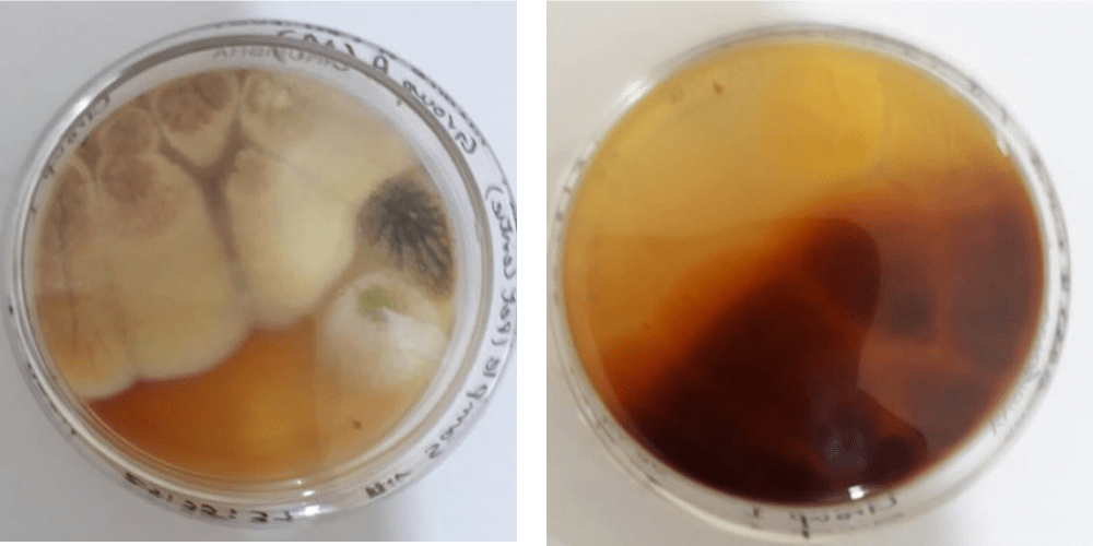
Aspergillus niger
It causes a disease called black mold on certain fruits and vegetables such as grapes, apricots, onions, and peanuts etc.
Macroscopic characters:
- Obverse character: black in color
- Reverse character: white or pale in colour
- Texture: initially hairy that turns velvety after 10 days.
- Margins: circular margins
- Top view: color of a colony from the top appear dense
Microscopic characters:
- Its hyphae is hyaline and septate.
- Asexual conidiophores are long and globose at the tip. They are generally smooth and colorless.
- Spores are globose and have conspicuous ridges or spines not arranged in row.
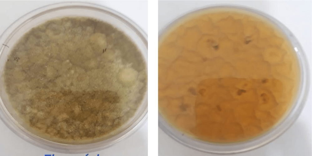
Aspergillus terrus:
The filamentous fungus was isolated from a bronchial washing from one of the patients.
Macroscopic characters:
- Obverse character: characteristic buff or cinnamon-brown colored.
- Reverse character: yellow to beige-brown.
- Texture: powdery/flour like/crumbly due to the presence of conidia
- Margins: regular circular margins.
- Top view: the color is olive green which is white at middle.
Microscopic characters:
- Conidiophores are smooth and hyaline.
- Conidia are small, globose shaped, and smooth walled and can vary from light yellow to hyaline.
- Asexual spores are directly produced on hyphae that are larger than phial conidia.
Mucor:
Microbial genus commonly found in soil, digestive system, plant surfaces etc.
Macroscopic characters:
- Obverse character: new growth is white in color but turns greyish brown with aging.
- Reverse character: pale white.
- Texture: woolly growth resembling cotton-candy like.
- Margins: entire colony is highly dense that completely covers the plate.
Microscopic characters:
- Hyphae are non-septate or sparsely septate.
- Arthrospores are present at end of hyphae and few chlamydospores may also be present.
- Apophysis, rhizoid and stolon are absent.
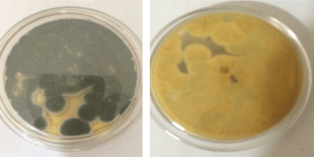
Aspergillus flavus:
It is a saprotrophic and pathogenic fungus with a cosmopolitan distribution.
Macroscopic characters:
- Obverse character: greenish yellow to olive color.
- Reverse character: cream to tan to yellowish on SDA agar.
- Texture: woolly tufts.
- Margins: regular circular margins.
- Top view: dark olive green
Microscopic characters:
- Its hyphae is hyaline and septate.
- Asexual conidiophores are long and globose at the tip. They are generally smooth and colorless.
- Spores are globose and have conspicuous ridges or spines not arranged in rows.
Aspergillus fumigatus
It is one of the most common Aspergillus species causing immunodeficiency.
Macroscopic characters:
It is a common culture contaminant.
- Obverse side: dull bluish green.
- Margins: rough circular and white margins.
- Texture: initially hairy that becomes dry mat after 2 weeks on maturation.
- Reverse side: creamy white and wrinkled colony from the reverse.
Microscopic characters:
- Hyphae are septate and unbranched conidiophores that arise from a specialized foot cell.
- The conidiophore is enlarged at the tip, forming a swollen vesicle. Vesicles are completely or partially covered with flask-shaped phialides.
- Phialides may be supported by a cell known as medulla. Phialides produce chains of mostly round, sometimes rough, conidia.
References
Salvamani, S., & Nawawi, N. M. (2014). Macroscopic and microscopic approaches for identification of fungi from plant soil of Cameron Highlands. Bioremediation Science and Technology Research, 2(1), 14–18. https://doi.org/10.54987/bstr.v2i1.68
Sibanda, Angela & Ruzvidzo, Oziniel & Ignatious, Ncube & Ncube, Thembekile. (2019). Diversity of cellulase- and xylanase-producing filamentous fungi from termite mounds. Journal of Yeast and Fungal Research. 10. 15-29. 10.5897/JYFR2019.0189.
https://www.anbg.gov.au/fungi/macroscopic.html
Frequently asked questions
What is the microscopic characteristic of fungi?
The microscopic characteristic of fungi refers to the features of fungi that can only be observed with the aid of a microscope, such as spore shape, size, color, and texture, as well as the presence or absence of certain structures like septa, hyphae, and fruiting bodies.
What are the macroscopic Characteristics of fungi?
The macroscopic characteristics of fungi refers to the features of fungi that can be observed with the naked eye or with the aid of a hand lens or dissecting microscope, such as colony color, texture, shape, and growth pattern.