
Introduction
S. typhi overview
Salmonella typhi is a well-known human pathogen that is responsible for typhoid fever. It is a Gram-negative bacterium that is a member of the Enterobacteriaceae family. This particular bacterium distinguishes itself by targeting the human gastrointestinal tract with specificity.
The result of a Salmonella typhi infection is typhoid fever, which is a systemic sickness affecting the digestive system. It is spread by consuming tainted food or drink, which gives the bacteria the opportunity to proliferate and grow inside the cells of the host. One of Salmonella typhi‘s unique characteristics is its ability to evade the immune system and take up residence in macrophages.
Salmonella typhi is a resilient organism that can survive in a variety of situations and adapt to a wide range of habitats. Although many types of Salmonella are harmless, Salmonella typhi in particular poses a threat to public health because of its connection to epidemics of typhoid
Isolation Techniques
Sample Collection
Thorough sample collection is essential for the isolation of Salmonella typhi. Different kinds of samples provide information on the prevalence and possible sources of this harmful bacteria.
- Water: Lakes and rivers, among other bodies of water, are important sites for sampling. Salmonella typhi can reveal fecal contamination and the possibility of waterborne transmission when it is found in water sources.
- Food: When it comes to foodborne diseases, food samples are essential for determining the possible origins of Salmonella typhi. Unpasteurized dairy products, tainted vegetables, and raw or undercooked meat are common sources of the bacteria.
- Clinical specimens: In clinical settings, stool and urine samples from patients displaying signs of upper respiratory tract infections or gastrointestinal disorders are extremely important. These specimens give medical personnel a clear connection between Salmonella typhi-related symptoms and infections, facilitating quick diagnosis and treatment.
Enrichment Media
To cultivate and support the growth of Salmonella typhi, specific enrichment media are employed, fostering an environment conducive to the proliferation of this pathogenic bacterium. The following enrichment media are commonly utilized:
- Selenite Broth
- Rappaport-Vassiliadis (RV) Broth
- Tetrathionate Broth
- Muller-Kauffmann Tetrathionate-Novobiocin (MKTTn) Broth
Salmonella typhi, when cultivated in these enrichment media, typically exhibits robust growth, resulting in distinctive characteristics such as dense, turbid cultures. These media play a crucial role in the isolation and subsequent identification of Salmonella typhi in various samples.
Isolation on Solid Media
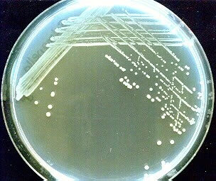
1. Nutrient Agar
While Salmonella typhi is not selective for specific nutrient media like MacConkey Agar, it can grow on general-purpose nutrient agar with the following characteristics:
Colony Morphology:
- Color: Salmonella typhi colonies often appear creamy or white on N agar.
- Appearance: Colonies may exhibit different morphologies, such as convex or raised.
- Size: Variable, ranging from small to large colonies.
- Texture: Texture can vary, depending on the strain.
Growth Patterns:
- Nutrient Utilization: Positive. Salmonella typhi can utilize the nutrients present in general-purpose nutrient agar for growth.
2. Xylose Lysine Deoxycholate (XLD) Agar
On Xylose Lysine Deoxycholate (XLD) Agar, Salmonella typhi displays distinct colony characteristics and growth features:
Colony Morphology:
- Color: Salmonella typhi colonies appear red with a black center.
- Appearance: Flat, moist, and translucent colonies.
- Size: Medium to large-sized colonies.
- Texture: Smooth and glistening.
Growth Patterns:
- H2S Production: Positive. Salmonella typhi produces hydrogen sulfide (H2S), leading to the formation of black precipitates or colonies in the agar.
- Lysine Decarboxylation: Positive. The utilization of lysine results in alkaline products, leading to the formation of red colonies.
- Xylose Fermentation: Positive. Salmonella typhi ferments xylose, contributing to the red coloration of colonies.
3. Hektoen Enteric (HE) Agar
Hektoen Enteric (HE) Agar supports the selective growth and identification of Salmonella typhi with the following characteristics:
Colony Morphology:
- Color: Salmonella typhi colonies exhibit blue-green to blue-black coloration.
- Appearance: Convex, moist, and slightly translucent colonies.
- Size: Medium-sized colonies.
- Texture: Smooth with an entire edge.
Growth Patterns
- Lactose Fermentation: Negative. Salmonella typhi does not ferment lactose, contributing to the lack of color change in the agar.
- Sulfur Reduction: Positive. Production of hydrogen sulfide (H2S) leads to a blackening of colonies.
- Fermentation of Sucrose: Negative. Salmonella typhi does not ferment sucrose.
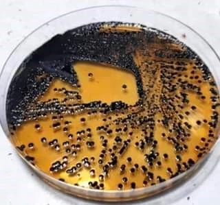
4. Bismuth Sulfite Agar
Bismuth Sulfite Agar is instrumental in isolating and identifying Salmonella typhi, showcasing the following characteristics:
Colony Morphology
- Color: Salmonella typhi colonies appear black.
- Appearance: Moist and opaque colonies.
- Size: Medium-sized colonies.
- Texture: Smooth with a shiny surface.
Growth Patterns
- Hydrogen Sulfide (H2S) Production: Positive. Salmonella typhi produces hydrogen sulfide, resulting in the blackening of colonies.
- Bismuth Sulfite Reduction: Positive. Reduction of bismuth sulfite contributes to the black coloration of colonies.
5. Salmonella Shigella (SS) Agar
On Salmonella Shigella (SS) Agar, Salmonella typhi exhibits distinctive characteristics:
Colony Morphology:
- Color: Salmonella typhi colonies appear colorless or pale.
- Appearance: Flat and translucent colonies.
- Size: Small to medium-sized colonies.
- Texture: Smooth with an entire edge.
Growth Patterns:
- Lactose Fermentation: Negative. Salmonella typhi does not ferment lactose, resulting in colorless colonies.
- H2S Production: Positive. Production of hydrogen sulfide leads to blackening of colonies.
6. MacConkey Agar
On MacConkey Agar, Salmonella typhi demonstrates distinctive colony characteristics and growth patterns:
Colony Morphology:
- Color: Salmonella typhi colonies appear colorless or pale.
- Appearance: Small, moist, and translucent colonies.
- Texture: Smooth with an entire edge.
Growth Patterns:
- Lactose Fermentation: Negative. Generally, Salmonella typhi does not ferment lactose, resulting in the absence of color change in the agar. However some strains are lactose fermenters and appear as pink colonies on MacConkey Agar.
- Colonial Characteristics: Non-lactose-fermenting colonies remain colorless against the pink background of the agar.
Microscopic Examination
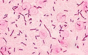
1. Gram Staining
- S. typhi, being a Gram-negative bacterium, appears pink/red under the microscope after Gram staining.
2. Morphological Characteristics Under the Microscope
- S. typhi is typically characterized by its small, rod-shaped or bacillus morphology when observed under the microscope.
- Generally, most of the strains of S. typhi are non-motile.
- However, some strains of S. typhi is possess peritrichous flagella and thus shows swimming or swarming motility these strains may also forms biofilms.
- Microscopic examination facilitates the estimation of cell size, with S. typhi cells typically measuring around 2–5 microns long by 0.5–1.5 microns wide.
Biochemical Tests
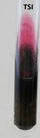
1. Triple Sugar Iron (TSI) Agar Test
- Result: Alkaline/Alkaline reaction, no gas, and hydrogen sulfide (H2S) production.
- Indication: Slant and butt both remain alkaline. No color change in the medium.
2. Citrate Utilization Test
- Result: Citrate-negative; no utilization of citrate as a sole carbon source.
- Indication: No color change in the medium.
3. Urease Test
- Result: Urease-negative; no hydrolysis of urea to produce ammonia.
- Indication: No color change in the urea agar.
4. Phenylalanine Deaminase Test
- Result: Phenylalanine deaminase-negative; no deamination of phenylalanine to phenylpyruvic acid.
- Indication: No green color development.
5. Sulfur-Indole-Motility (SIM) Medium Test
The SIM (Sulfide, Indole, Motility) test is often used to assess three key characteristics of Salmonella typhi:
- Sulfide Production: Positive (blackening of the medium)
- Indole Production: Negative – no indole production from tryptophan (Absence of purple ring)
- Motility: Negative (Limited or no growth away from the stab line)
6. Lysine Decarboxylase Test
- Result: Lysine decarboxylase-positive; production of alkaline products.
- Indication: Purple color change in the medium, indicating Salmonella typhi growth.
7. Oxidase Test
- Result: Typically, oxidase-negative.
- Indication: No color change on the oxidase test strip.
8. Catalase Test
- Result: Catalase-positive; production of oxygen from hydrogen peroxide.
- Indication: Bubbling or effervescence when hydrogen peroxide is added.
9. Widal Test:
- Result: Positive result indicated by agglutination of O and H antigens.
- Indication: The presence of antibodies against Salmonella typhi antigens leads to visible clumping or agglutination, suggesting a current or past infection with the bacterium.
- The intensity of agglutination, measured by titer levels, aids in the diagnosis of typhoid fever

Biochemical profile
| BASIC CHARACTERISTICS | PROPERTIES (S. TYPHI) |
| Shape | Cocci |
| Gram Staining | Positive (+ve) |
| Spore | Non-Sporing |
| Capsule | Non-Capsulated |
| Pigment | Mostly Positive (+ve) |
| Oxidase | Negative (-ve) |
| Catalase | Positive (+ve) |
| Coagulase | Positive (+ve) |
| Citrate | Positive (+ve) |
| Urease | Positive (+ve) |
| VP (Voges Proskauer) | Positive (+ve) |
| MR (Methyl Red) | Positive (+ve) |
| Nitrate Reduction | Positive (+ve) |
| Indole | Negative (-ve) |
| H2S | Negative (-ve) |
| Gas | Negative (-ve) |
| Motility | Negative (-ve) |
| Gelatin Hydrolysis | Positive (+ve) |
| Hemolysis | Positive (+ve)- Beta |
| OF (Oxidative-Fermentative) | Fermentative |
| PYR | Negative (-ve) |
Sugar Fermentation profile
| SUGARS | FERMENTATION RESULTS |
| Glucose | Positive (+ve) |
| Fructose | Positive (+ve) |
| Sucrose | Positive (+ve) |
| Lactose | Positive (+ve) |
| Maltose | Positive (+ve) |
| Galactose | Positive (+ve) |
| Mannose | Positive (+ve) |
| Mannitol | Positive (+ve) |
| DNase | Positive (+ve) |
| Ribose | Positive (+ve) |
| Arabinose | Negative (-ve) |
| Cellobiose | Negative (-ve) |
| Trehalose | Positive (+ve) |
| Xylose | Negative (-ve) |
| Raffinose | Negative (-ve) |
| Salicin | Negative (-ve) |
Enzymatic reactions profile
| ENZYMES | DEGRADATION REACTIONS |
| Hyaluronidase | Positive (+ve) |
| Lipase | Positive (+ve) |
| Ornithine Decarboxylase | Negative (-ve) |
| Acetoin Production | Positive (+ve) |
| Alkaline Phosphatase | Positive (+ve) |
| Arginine Dehydrolase | Positive (+ve) |
Molecular Techniques for E. coli Identification
1. Polymerase Chain Reaction (PCR)
- Efficient amplification of specific DNA sequences for S. typhi identification.
- Utilizes target genes to ensure accurate and reliable results.
Target Genes for S. typhi Identification
- ompC Gene (Outer Membrane Porin C)
- viaB Gene (Vi Antigen Biosynthesis)
- tyv Gene (Tyvelose Pathogenicity Island)
- phoE Gene (Outer Membrane Protein E)
2. DNA Sequencing:
- Determination of the nucleotide sequence of S. typhi DNA.
- High accuracy in identifying specific genetic markers.
3. Real-time PCR:
- Quantitative measurement of DNA amplification in real-time.
- Allows for monitoring the progress of the reaction.
4. Multiplex PCR:
- Simultaneous amplification of multiple S. typhi target genes.
- Increased efficiency in identifying the bacterium.
5. Pulsed-Field Gel Electrophoresis (PFGE):
- Separation of large DNA fragments for genetic fingerprinting.
- Useful in epidemiological studies.
6. Fluorescent In Situ Hybridization (FISH):
- Visualization of specific S. typhi DNA sequences within intact cells.
- Fluorescent probes enhance specificity.
References
- Ruiz, J., Núñez, M. L., Lorente, I., Pérez, J., Simarro, E., & Gómez, J. (1996). Performance of six culture media for isolation of Salmonella species from stool samples. European journal of clinical microbiology & infectious diseases : official publication of the European Society of Clinical Microbiology, 15(12), 922–926. https://doi.org/10.1007/BF01690509
- Murray, P. R., Rosenthal, K. S., Pfaller, M. A., & Goldstein, E. J. (2019). Medical Microbiology. Elsevier.
- Forbes, B. A., Sahm, D. F., & Weissfeld, A. S. (2007). Bailey & Scott’s Diagnostic Microbiology (12th ed.). Mosby.