
Introduction
Microbiologists use many different types of microscopes to do their work. The most common ones are bright-field, dark-field, phase-contrast, and fluorescence microscopes.
Magnification in a microscope depends on the kinds of lenses it has and how many of them it has. There are two types of microscopes based on how many lenses they have. The simple light microscope and the compound light microscope. Modern microscopes are all compound microscopes. The lenses are lined up so that they can bend light in a way that makes the image larger.
A working light microscope needs to be able to focus a beam of light through a very small and clear sample to make an image. The image is then put through one or two lenses to make it bigger so that you can see it easily. Because the specimen is clear, light can easily and quickly pass through it. Specimens can be made up of bacteria, cells, and other tiny living things.
light microscopes use a glass lens to see an image, and the amount of magnification depends on how well the lens can bend light and focus it on the specimen, making an image. When a ray of light goes from one medium to another, it bends at the boundary between the two. This is called refraction. The refractive index shows how much light bends when it passes through it. and refractive index is a measure of how much a substance slows down the speed of light. The refractive indices of the two materials that make up the interface determine which way the light bends and how much it bends.
A brief history of light microscope
- Late 16th century: Dutch spectacle maker Zacharias Janssen invents the compound microscope by placing two lenses in a tube, but the image produced was blurry.
- 17th century: Robert Hooke observes and describes cells using a compound microscope, while Antonie van Leeuwenhoek develops a simple microscope that allowed him to observe microorganisms with great clarity.
- 18th century: Improvements are made to the lenses and illumination systems used in light microscopes, resulting in clearer images.
- 19th century: The development of achromatic lenses and improvements to the stage and focus mechanisms greatly improve the quality of images produced by light microscopes.
- 20th century: The invention of electron microscopy leads to a temporary decline in the use of light microscopes, but advances in technology lead to the development of confocal microscopy, super-resolution microscopy, and other forms of high-resolution light microscopy.
- Today: Light microscopy continues to be a vital tool in many fields of science and medicine, with new techniques and technologies constantly being developed to further improve the capabilities of these instruments.
also read Electron Microscope: Its definition, types, and advantages
How does a light microscope work?
When a light goes through a medium with a lower refractive index, like glass to air, it usually moves faster and bends away from the normal. When a light goes through a medium with a higher refractive index, like air to glass, it usually moves slower and bends towards the normal, perpendicular to the surface.
If you put something between the water and the air, like a prism, in this case, the light will bend at an angle. This is how microscopic lenses work: they bend light at an angle. When the light hits the convex lens, the light rays come together at a single point called the focal point (F-point). The focal length is the distance between the center of the lens and the focal point. See figure 1
The power of the lenses in a microscope is already set. The power of a lens is directly related to its focal length, so lenses with a short focal length make things look bigger than lenses with a long focal length.
Microscopy is based on a single factor called “resolution.” The ability of a lens to distinguish small objects that are close to each other is known as resolution. The resolution of a light microscope is based on the numerical aperture of its lens system and the wavelength of the light it uses. The ability of a microscope objective to gather light and resolve fine specimen detail while working at a fixed object (or specimen) distance is known as a numerical aperture.
Using the wavelength of the light that hit the specimen and the numerical aperture. You can use the Abbe equation to figure out the minimum distance (d) between two objects that makes them look like two different things. For example, d=0.5 /n sin.
Types of light microscopy
There are 3 types of modern light microscopes which include
- Bright field Light Microscope
- Dark-Field Light Microscope
- Phase Contrast Microscope
- Fluorescence Microscope
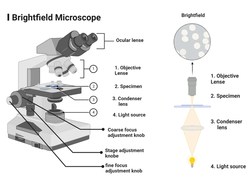
Bright-field light microscope
In microbiology labs, the bright field microscope is frequently used to examine both stained and unstained specimens. Because it creates a dark image against a brighter background, the instrument is known as a bright-field microscope.
Parts of Bright field light microscope
- It is made up of a strong metal body or stand with a base and an arm to which the other components are attached. The base carries a light source, which could be a mirror or an electric illuminator. The fine and coarse adjustment knobs, which can move the stage or the nosepiece to focus the image, are two focusing knobs that are found on the arm.
Condenser
- The stage is About halfway up the arm. It has either simple slide clips or a mechanical stage clip to hold the slides. With the knobs on a mechanical stage, the operator can move a slide smoothly while looking at it. The substage condenser, also called a “condenser,” is attached to the stage or below it. It focuses a cone of light on the slide. In simple microscopes, its position is often fixed, but in more complex ones, it can be moved up and down.
- The curved top part of the arm holds the body assembly, which has a nosepiece and one or more eyepieces or ocular lenses attached. Binocular microscopes are more advanced microscopes that have eyepieces for both eyes. The body assembly has a series of mirrors and prisms that allow the barrel that holds the eyepiece to be tilted so that viewing is easier.
What is Parfocal?
- The nosepiece holds three to five objective lenses with different levels of magnification, and it can be rotated so that any objective can be placed under the body assembly. In an ideal case, a microscope should be parfocal, which means that when the objectives are changed, the image should stay in focus
- When you look at a specimen with a compound microscope, the objective and ocular lenses work together to make the image you see. The objective lens focuses the light from the illuminated specimen, making a larger image inside the microscope.
- During visualization, the objective lens stays parfocal, which means that when the objective lens is changed, the image stays in focus. The objective lens is an important part of the microscope that determines how the image is focused on the condenser to make a large, clear image inside the microscope. The eyepiece then magnifies this image even more to make a primary image. ( see Figure 1 )
Magnification of Bright field light microscope
The clear image of the specimen that you can see when you look at it through a microscope is called the “virtual image.” To figure out the magnification, you need to multiply the objective magnification by the eyepiece objective magnification. The magnification is standard, which means that it is neither too high nor too low. Depending on how powerful the magnification of lenses is. the magnification will range from 40X to 100X.
- Calculation of magnification = Magnification of objective lens/magnification of the eyepiece lens
Resolution of Bright field light microscope
The objective lens is very important because it not only makes the image larger but also makes it clear to see it. This is called resolution. Prescott says that a lens’s resolution is its ability to tell the difference between small things that are close together.
Even though the eyepiece magnifies the image at the end of the viewing, its range is smaller than that of the objective lens. The eyepiece’s range is 8X-12X (10X standard), while the objective lens’ range is 40X-100X. The objective lens is responsible for most of the microscope’s magnification and resolution.
also read: Bacterial Morphology: size, shape, and arrangements
Dark field Microscope
Using this method, you can see living, unstained cells. This depends on how the specimen is illuminated. When a hollow cone beam of light is transmitted to the specimen, light rays that are not reflected or refracted do not pass through the objectives, but light rays that are reflected or refracted do pass through the objectives and make an image on the specimen.
This makes the area around the specimen look dark, while the specimen itself looks bright. This is possible because the background is dark, which is where the term “dark field” comes from.
Fluorescent Microscope
In a fluorescent microscope, the specimen emits light. By adding a molecule of dye to the sample. This dye molecule gets excited when it gets light energy, so it emits any energy it has stored as light. The excited molecule gives off light energy, which has a longer wavelength than the light it gives off. Most of the time, the dye molecule is a fluorochrome, which glows when exposed to a certain wavelength of light.
The principle is that the fluorescent microscope exposes the specimen to ultraviolet or blue light, which makes an image of the sample that comes from the fluorescent light. They have a mercury vapour arc lamp that makes a strong beam of light that goes through an exciter filter. At the objective, the fluorochrome-labeled image is made by sending a certain wavelength through the exciter filter to the fluorochrome-stained specimen.
Applications of Light Microscopy
Light microscopy is a powerful tool for examining the structure and function of biological and non-biological specimens. Here are the most common applications of light microscopy:
Observing cells and tissues
One of the most important applications of light microscopy is observing cells and tissues. Biologists use light microscopy to observe the morphology, structure, and behavior of cells and tissues. This type of microscopy is used to study the physiology of cells, the development of tissues, and the pathology of diseases.
Microbial identification
Microorganisms are too small to be seen with the naked eye, but they can be observed and identified using light microscopy. Microbial identification is an important application of light microscopy, and it is used in microbiology to study the morphology and behavior of microorganisms. This type of microscopy is used to identify pathogens that cause diseases and to study the ecology of microorganisms in different environments.
Medical diagnosis
Light microscopy is used in medical diagnosis to observe cells and tissues from patients. Medical professionals use light microscopy to diagnose diseases such as cancer, infections, and autoimmune disorders. This type of microscopy is also used in hematology to study blood cells and to diagnose blood disorders such as anemia and leukemia.
Forensic investigation
Light microscopy is used in forensic investigation to examine physical evidence such as hair, fibers, and soil samples. Forensic scientists use light microscopy to compare the physical characteristics of the evidence with those of known samples to identify the source of the evidence. This type of microscopy is also used to study the morphology and behavior of insects and other small organisms that can provide clues about the time and location of a crime.
Recent Advances in Light Microscopy
Light microscopy has advanced rapidly in recent years, and new techniques have been developed that allow scientists to observe biological and non-biological specimens in unprecedented detail. Here are some of the most recent advances in light microscopy:
Super-Resolution Microscopy
Super-resolution microscopy is a technique that allows scientists to observe structures that are smaller than the diffraction limit of light. This type of microscopy uses fluorescent dyes that emit light when excited by a laser beam. The fluorescent signals are captured and analyzed to create an image with higher resolution than traditional light microscopy. Super-resolution microscopy has revolutionized the study of biological structures and has provided new insights into the organization and function of cells.
Digital Holographic Microscopy
Digital holographic microscopy is a technique that uses holography to create three-dimensional images of specimens. This type of microscopy uses a laser beam to create a hologram of the specimen, which is then captured by a camera. The hologram is reconstructed using computer software to create a 3D image of the specimen. Digital holographic microscopy has many applications in biology, including the study of cell motility and the visualization of subcellular structures.
Single-Molecule Imaging
Single-molecule imaging is a technique that allows scientists to observe the behavior of individual molecules in real time. This type of microscopy uses fluorescent dyes that are attached to molecules of interest. The fluorescent signals are captured and analyzed to create a movie of the movement and interactions of the molecules. Single-molecule imaging has revolutionized the study of molecular biology and has provided new insights into the function and regulation of biological processes.
Conclusion
In conclusion, light microscopy is a crucial tool in the study of biology, medicine, and many other fields of science. It has played a vital role in advancing our understanding of the natural world, allowing us to observe and study cells, tissues, microorganisms, and other small objects in great detail.
From its humble beginnings in the late 16th century to the high-tech instruments of today, the light microscope has undergone significant advancements that have greatly improved its capabilities and resolution. With the recent developments in confocal microscopy, super-resolution microscopy, and other advanced techniques, the possibilities for research and discovery using light microscopy are endless. Overall, light microscopy remains an indispensable tool for scientists and researchers around the world, enabling us to see and explore the hidden world of microcosms.
References
- Tortora, G. J., Funke, B. R., & Case, C. L. (2021). Microbiology: An introduction. Pearson Education Limited.
- Willey, J. M., Sandman, K. M., Wood, D. H., & Prescott, L. M. (2019). Prescott’s microbiology (11th ed.). McGraw Hill.
- Hardin, J., Bertoni, G., & Becker, W. M. (2018). Becker’s world of the cell. Pearson.
- Cappuccino J.G. and Sherman N. 2008. Microbiology: A Laboratory Manual, 8th ed. Pearson Benjamin Cummings, San Francisco, CA, USA.
- Clinical Microbiology Procedures Handbook, Fourth Edition. (2016). In Clinical Microbiology Procedures Handbook, Fourth Edition. American Society of Microbiology. https://doi.org/10.1128/9781555818814