The most dynamic component of a prokaryotic cell is the plasma membrane, sometimes referred to as the cytoplasmic membrane. Its primary role is as a selective permeability barrier that controls how quickly chemicals can enter and leave the cell. Since it confines the molecules of life in a single unit and isolates the cell from its surroundings, the plasma membrane is the distinguishing feature of a cell.
Water and uncharged molecules up to an MW of around 100 Daltons can flow across the bacterial membrane, but larger molecules or any charged compounds cannot unless they are transported through specialized membrane transport mechanisms and processes. Bacterial membranes are made up of 60% protein and 40% phospholipid.
The phospholipids, which naturally form a bilayer in aqueous settings are amphiphilic molecules with two nonpolar hydrophobic fatty acid tails joined by an ester link to a polar hydrophilic glycerol “head”. Most membrane functions are performed by a variety of structural and enzymatic proteins dispersed across the bilayer.
Explain the plasma cell membrane structure.
Phospholipids:
The head and the two tails of a phospholipid make up the compound which are both significant components. A phosphate molecule that serves as the head is attracted to water (hydrophilic). Fatty acids (chains of carbon atoms) that make up the two tails are repellent to water (hydrophobic). On both the inside and exterior of the cell, the cell membrane is exposed to water mixed with electrolytes and other substances.
Because of their hydrophilic and hydrophobic properties, phospholipids cluster into two layers when cellular membranes are formed. Because they are hydrophobic, the phosphate tails in between the layers of heads avoid contact with the water while the heads in each layer face the aqueous or watery environment on either side. This effective assembling capability is known as “self-assembly” in biology.
Proteins:
Transmembrane proteins are a subset of proteins that are located in the lipids that make up the membrane and create channels, apertures, or gates that open passage for molecules that would otherwise be unable to enter the cell. The cell regulates the flow of these chemicals into and out of the cell in this manner. Numerous other processes, such as cell signaling, cell recognition, and enzyme activity, depend on proteins in the cell membrane.
Carbohydrates:
The plasma membrane contains carbohydrates as well; particularly, most of the carbohydrates there are found as components of glycoproteins, which are created when a carbohydrate binds to a protein. Glycoproteins are involved in cell adhesion, which is how cells bind to one another, as well as other cell-cell interactions.
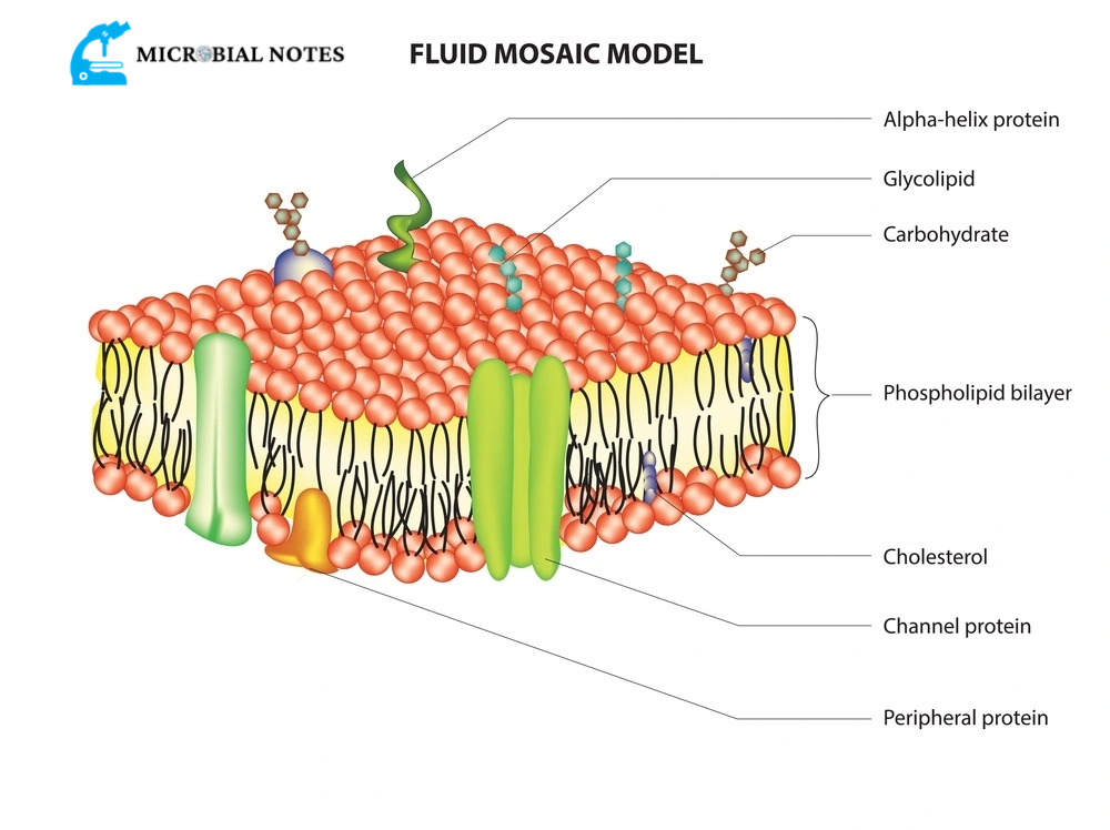
Fluid mosaic model:
Different facts on the composition of functional cell membranes are explained by the fluid mosaic hypothesis. According to this biological model, protein molecules are embedded within a lipid bilayer, which is a two molecules thick layer made mostly of amphipathic phospholipids. The membrane’s fluidity and elasticity are provided by the phospholipid bilayer. The cell membrane also contains trace amounts of carbohydrates.
The cell membrane is portrayed in the biological model as a two-dimensional liquid that prevents the lateral diffusion of membrane components, which was developed in 1972 by Seymour Jonathan Singer and Garth L. Nicolson. Such domains are identified by specific lipid and protein cocoon-rich areas of the membrane that encourage the development of lipid rafts or protein and glycoprotein complexes.
The interaction of the lipid membrane with the cytoskeleton filaments and the extracellular matrix via membrane proteins is another approach to describing membrane domains. The current model covers several biological processes, including cell-cell communication, apoptosis, cell division, membrane budding, and cell fusion. The most palatable plasma membrane model is the fluid mosaic one. Its major job is to keep the cell’s interior from the outside world.
Difference between the prokaryotic plasma membrane and eukaryotic plasma membrane
Although bacterial membranes share structural similarities with those of eukaryotes, they are made of saturated or monounsaturated fatty acids (rarely polyunsaturated fatty acids) and often lack sterols. Archaeal lipids are saturated, branched, repeating isoprenoid subunits that attach to glycerol via an ether linkage. While an ester linkage found is in glycerides of eukaryotic and bacterial membrane lipids.
Also, read prokaryotes vs eukaryotes its characteristics and differences
Although the membranes of Archaea form bilayers that are functionally equivalent to those of bacteria. It is believed that the structure of archaeal membranes represents a survival strategy for them in harsh conditions. In comparison to prokaryotes, the eukaryotic plasma membrane contains a broader range of fatty acids and more negatively charged phospholipids. Prokaryotes lack cholesterol, but it is present in the plasma membrane of eukaryotes. Prokaryotes, however, contain hopanoids (another derivative of sterol).
Functions:
Because prokaryotes lack intracellular organelles for activities like respiration, photosynthesis, or secretion, the plasma membrane takes on these roles for the cell and serves a multitude of purposes in biosynthesis and energy production. The prokaryotic membrane, for instance, houses the electron transport system that connects aerobic respiration and ATP production.
The membrane contains photosynthetic chromophores, which convert light energy into chemical energy. As a result, the prokaryotic plasma membrane serves as the site of oxidative phosphorylation and photophosphorylation, like eukaryotic mitochondria and chloroplasts which serve these purposes. Prokaryotic membranes may also contain sensing proteins that measure molecule concentrations in the environment or binding proteins that transport signals to genetic and metabolic machinery in the cytoplasm.
They also have transport proteins that selectively mediate the passage of substances into and out of the cell. Enzymes found in membranes are also engaged in a wide range of metabolic processes, including the production of cell walls, the development of septa, the synthesis of membranes, DNA replication, CO2 fixation, and ammonia oxidation.
Purposes:
The main purpose of the prokaryotic cell membrane is as follows:
- Permeability or osmotic barriers
- Where the transport networks for various solutes are located (nutrients and ions)
- Energy-producing processes that include the electron transport chains of the respiratory and photosynthetic systems, the creation of the proton motive force, and the transmembrane, ATP-synthesizing ATPase
- The production of membrane lipids (including lipopolysaccharide in Gram-negative cells)
- Production of murein (cell wall peptidoglycan)
- Protein synthesis and secretion from extracellular space
- DNA replication and segregation are coordinated with cell division, septum creation, and DNA replication
- Chemotaxis (both movement per se and sensing functions) (both motility per se and sensing functions)
- Location of the specific enzyme system
References
Hardin, J., Bertoni, G., & Becker, W. M. (2018). Becker’s world of the cell. Pearson.