History of peroxisomes:
De Duve et al. extracted microbodies from the rat liver and termed them peroxisomes in 1965. They were membrane-bound organelles that included a variety of oxidases that produce H2O2 and catalase, degrading H2O2. In 1976, the fatty acid -oxidation mechanism in rat liver peroxisomes was discovered.
Peroxisomes lack DNA and are surrounded by a single membrane, unlike mitochondria and chloroplasts, although it has been proposed that they are symbiogenetic and descended from an anaerobic prokaryote that produces hydrogen in captivity.
Define peroxisomes.
In most eukaryotic cells, peroxisomes are membrane-bound organelles that play a key role in lipid metabolism and the transformation of reactive oxygen species like hydrogen peroxide into safer molecules like oxygen and water.
Due to their high energy density, fats are useful energy storage molecules. One gram of fat is converted into substantially more ATP when it is oxidized than it is when it is converted into carbs or proteins.
Lipids are also very helpful chemicals for dividing cells into membrane-bound compartments or separating the cytoplasm from the external environment. However, they are difficult to process in an aqueous cellular environment due to their lipophilic biochemistry. These hydrophobic compounds are metabolized in peroxisomes, which are biological cells.
Explain the structure of peroxisomes.
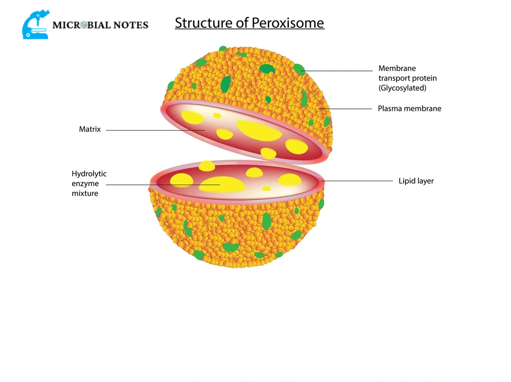
Depending on the cell’s energy requirements, peroxisomes’ size, shape, and number can change. A growth medium-high in carbohydrates causes the peroxisomes in yeast cells to shrivel. On the other hand, pollutants or a diet high in lipids can make them more numerous and larger.
The phospholipid bilayer that makes up these organelles contains numerous membrane-bound proteins, including those that function as protein transporters and translocators. In contrast to lysosomes that sprout from the endoplasmic reticulum, the enzymes involved in detoxification and lipid metabolism are generated on free ribosomes in the cytoplasm and are selectively transported into peroxisomes (ER).
However, there is also some evidence connecting peroxisome-resident enzymes to ER-mediated protein synthesis. In general, one of two signal sequences is included in enzymes and proteins that are intended for the peroxisome. That is, there are brief intervals of a few amino acids that identify the protein’s subcellular location. The Peroxisome Targeting Sequence 1 (PTS1), which is an amino acid trimer, is the most popular signal sequence.
A serine residue, a lysine, and a leucine residue are located in the carboxy-terminal end of proteins that contain the PTS1 signal sequence. This signal sequence is present in a significant majority of peroxisomal proteins. Amino acid sequences upstream of this trimer are also required for PTS1 to operate properly. According to some sources, the C-terminal sequence should ideally be viewed as a length of 20 amino acids that are required for the peroxisomal transporter and translocator molecules to recognize the protein.
An alternative would be for a peroxisomal protein to have a 9-amino acid N-terminal signal sequence. This sequence consists of two dimers that are spaced apart by five amino acids. Leucine and arginine combine to form the first dimer, while histidine and leucine combine to form the second dimer. The single-letter amino acid coding for this signal sequence is RLx5HL.
There is some proof that there are uncharacterized additional internal sequences that target proteins for import into the peroxisome. Additionally, peroxisomes may have the appearance of a crystalloid core and contain a few enzymes in very high concentrations.
The smooth ER serves as the primary site of phospholipid synthesis for peroxisomes. Peroxisome can split into two organelles as it expands in size as a result of the entry of proteins and lipids.
Explain the function of peroxisomes.
Hydrogen peroxide metabolism:
Enzymes found in peroxisomes are involved in the metabolism of hydrogen peroxide (H202), a reactive oxygen species that are produced and eliminated by the body.
Fatty acid oxidation:
In animal cells, fatty acids are oxidized both in the mitochondria and the peroxisomes, but solely in the peroxisomes of yeasts and plants. The enzyme catalase breaks down the H202 that is produced in conjunction with oxidation. Significant metabolic energy is provided by this.
Lipid biosynthesis:
Both ER and peroxisomes are involved in cholesterol and dolichol synthesis. In the liver, cholesterol is used to create bile acids. A family of phospholipids called plasmalogens, which are crucial membrane constituents of heart and brain tissue, is synthesized by enzymes found in peroxisomes.
Plant seed germination:
Peroxisomes in seeds are in charge of converting fatty acids that have been stored therein into carbohydrates, which are essential for supplying energy and raw materials for the growth of germination plants.
Photorespiration:
Together with chloroplasts, peroxisomes in leaves, especially green ones, perform the photorespiration process.
Transformation of purines:
Purine, polyamine, and amino acid catabolism should be carried out, in particular by uric acid oxidase.
Bioluminescence:
Firefly peroxisomes contain the luciferase enzyme, which promotes bioluminescence and helps the flies find a mate or a meal.
What are the disorders related to peroxisome function?
Disorders resulting from impaired peroxisome function may be caused by flaws in peroxisome biogenesis, altered peroxisomal enzymes, or inoperable transporters that bind cytoplasmic proteins that contain PTS1 and PTS2. Rare genetic illnesses with delayed neuronal migration, myelin insufficiency, and poor brain development are the most severe of these. The skeletal system, liver, kidney, eyes, heart, and lungs are a few additional organs that are impacted.
These illnesses are typically brought on by mutations in the PEX genes, which are required for the biogenesis of organelles, including the creation of the subcellular membrane and the identification and import of cytoplasmic proteins into the organelle matrix. For instance, PEX2 creates the translocation channel for the import of matrix proteins, whereas PEX16 is involved in the production of peroxisomal membranes. On the other hand, the receptor for the PTS1 signal sequence is PEX5.
Defects in these proteins can result in the abnormal presence of phospholipids like plasmalogens in red blood cells as well as the buildup of long-chain fatty acids in blood plasma or urine.
What are peroxisomal enzymes?
At least 50 distinct enzymes, found in peroxisomes, participate in a wide range of metabolic pathways in various cell types. Originally, peroxisomes were referred to as organelles that perform oxidation processes that result in the generation of hydrogen peroxide.
- The matrix of peroxisomes contains over 60 recognized enzymes.
- They are in charge of performing the oxidation processes that result in the creation of hydrogen peroxide.
- The principal categories of enzymes are:
- Urate Oxygenase
- Deoxy D-amino acid oxidase
- Catalase
Metabolism of peroxisome:
Small molecules like sugar are able to pass through isolated peroxisomes. They frequently lose proteins that are normally included in the peroxisomal matrix during the isolation procedure. Peroxisomes are single membrane enclosed, hermetically sealed vesicles found in all living cells.
Explain peroxisomes in plants.
Peroxisomes are crucial to photosynthesis and seed germination in plants. Fat reserves are released during seed germination to fuel anabolic processes that result in the synthesis of carbohydrates. The glyoxalate cycle starts with -oxidation, which also results in the production of acetyl CoA. In leaves, peroxisomes recycle the byproducts of photorespiration to stop energy loss during photosynthetic carbon fixation.
For photosynthesis to occur, an essential enzyme known as ribulose-1,5-bisphosphate carboxylase/oxygenase (RuBisCO) that catalyzes the carboxylation of ribulose-1,5-bisphosphate is required (RuBP). This is the main chemical process that fixes carbon dioxide to create organic compounds. But as its name suggests, RuBisCO may also oxygenate RuBP by utilizing molecular oxygen and producing carbon dioxide, thereby undoing the overall effects of photosynthesis. The stomata close in order to stop transpiration when the plant is exposed to hot, dry surroundings, which is particularly true.
Phosphoglycolate, a 2-carbon molecule, is created when RuBisCO oxidizes RuBP. Peroxisomes take this in and oxidize it into glycine. After that, it moves back and forth between the mitochondria and peroxisomes where it goes through a number of changes before becoming a glycerate molecule that may be imported into chloroplasts to take part in the Calvin cycle for photosynthesis.
Lipid biosynthesis and detoxification:
Peroxisomes are the locations for a portion of the lipid biogenesis in animal cells, particularly for the unique phospholipids known as plasmalogens that build the myelin sheath in nerve fibers. Bile salt production also needs peroxisomes to take place.
In these organelles, about 25% of the alcohol we eat is converted to acetaldehyde. They make up a significant portion of kidney and liver cells because of their function in detoxifying and oxidizing a variety of chemicals, metabolic byproducts, and medications.
Comparison between peroxisome and other organelles:
Peroxisomes and other cellular organelles share some structural characteristics. At first, even lysosomes and peroxisomes could not be distinguished by microscopic analysis. The content of these two subcellular structures was then revealed by differential centrifugation. They comprise enzymes that are quite diverse from one another and have separate protein and lipid components.
In particular, catalase is present in peroxisomes to detoxify the hydrogen peroxide produced by the beta-oxidation of lipids. Lysosomal proteins are made in the rough ER, and vesicles with the right enzymes branch off to create the lysosome, which is another significant difference.
Peroxisomes resemble chloroplasts and mitochondria in some ways. On free ribosomes in the cytoplasm, these organelles’ majority of proteins are translated. Peroxisomes, in contrast to mitochondria and chloroplasts, do not have any genetic material or translational apparatus; as a result, their complete proteome is imported from the cytoplasm. Peroxisomes are also formed by a single lipid bilayer membrane as opposed to the double membranous structures of mitochondria and chloroplasts.

