Viruses are the smallest living creatures on the earth which is just a particle outside the host and deadly pathogens inside the host, nowadays the most crucial topic due its infection and damage to public health and the economy. Virus quantification assays are the best way to diagnose virus-host interaction and virus pathogenicity. One of the quantification methods is Infective dose 50 such as TCID50. Which is the amount of virus required to infect 50 percent of the healthy hosts like humans, animals and plants, etc
It depends upon the host you want to calculate ID 50 if you are using eggs that are called EID 50 and If you are using cell culture that is called TCID 50
What is TCID50?
Tissue culture infective dose 50 is the minimum amount of a virus to infect 50 percent of cells in cell culture. For measuring the generated data, there are several statistical methods (such as Reed–Muench Method or Spearman-Karber analysis). When insufficient experimental material or analysis expenses prevent lengthy repeat titer measurements, it is necessary to find out the inaccuracy of the methods. as compared to plaque assay which is a more accurate method but TCID 50 is more cost-effective and easy to perform because there are many viruses that cannot show plaques. Therefore, TCID50 is used extensively for a variety of applications in experimental and diagnostic virology.
Seeding of cells in 96 well plates
According to most of the articles, 1X10⁴ numbers cells are required to make confluency inappropriate on days. for example, If you add fewer numbers of cells it will take time to become 80 % confluent. and if you add a large number of cells it will become over confluent and the result may effect due to it. Alternatively, depending on the cell line needed for viral propagation, a different cell density can be necessary. Plates should be gently shaken back and forth and side to side to disseminate cells equally. After seeding the cells, let them continue to develop throughout the night. The next day, observe cells under a light microscope to check that they have attained above 80% confluency and are spread uniformly.
How to dilute virus?
The next step is the dilution of the virus, Dilutions of the sample containing the original virus should be made repeatedly at a ratio of 1:10.
- Take 10 tubes in a rack and label it 10-1 to 10-10
- Add 900ul of diluent( it may be DMEM or PBS) in each tube
- In 1st tube, add 100ul of virus sample and mix it with the vertex machine
- Then transfer 100ul from the 1st tube to the 2nd tube to make a dilution of 1:100 and again mix it well by vertex machine.
- Continually repeat the process to dilute the virus by a factor of 1:10, from 10-3 to 10-10.
Infection of the virus to the monolayer of cells
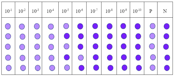
- Take 96 well plates containing 80 % confluent monolayer .label each column ( 8 wells) according to dilution 10-1 to 10-10
- The 11th column will be Positive control by adding 100ul of stock virus
- The 12th column will be negative control by adding 100ul of media.
- Add 100ul of 10-1 dilution of virus in each well of the 1st column
- Add 100ul of 10-2 dilution of virus in each well of the 2nd column
- Add 100ul of 10-3 dilutions of virus in each well of the 3rd column and so on till the 10th column
- Incubate at 37 C for 1 hour to settle down
- After incubation add 100ul of DMEM in all wells and incubate for 48 hours to show CPE
What is CPE?
A virus alters the structure by invading the host cell. This is called the cytopathic effect. The condition develops when the cell’s infection causes lysis or dies due to its inability to reproduce. Cytopathogenic viruses cause morphological alterations in their host cells. It all depends upon the types of cells and the types of viruses that are used to infect cells
The initial sign of viral infection or cytopathic impact is the rounding of cells. In the cytoplasm and nucleus of the host cell, inclusion bodies may be seen. Through optical or electron microscopy, they can be seen in the patients’ blood smears. Syncytia are a result of certain viral infections. These large masses in cytoplasm form when infected cells are fused. There are several nuclei in it.
Visualization and calculation of CPE
CPE can be visualized according to the type of virus and the types of cells used for the test. After incubation, you will observe each well under the microscope and check the positive and negative control as well for quality control and test validity. CPE should be present in the positive control and cells should remain intact in the negative control. Draw a table, and Observe 8 wells of each column and write a positive sign for CPE presence. According to Reed–Muench Method
| Dilution | Positive wells/total wells | Cumulative positive(A) | Comulative negative(B) | Infection rate A/(A+B) |
|---|---|---|---|---|
| 104 | 5/5 | 9 | 0 | 100 |
| 105 | 3/5 | 4 | 2 | 66.67 |
| 106 | 1/5 | 1 | 6 | 14.3 |
| 107 | 0/5 | 0 | 11 | 0 |
As Table 1 shows that tcid50 falls between log dilution of 10-5 and 10-6
Formula of TCID50
PD = 50% above positive-50/ 50% above positive- 50% below Positive
By using the above formula
PD = 66.67-50/66.7-14.3 = 0.318
TCID50 = 10^-5 + 0.318 = 10^-5.318
Hence 10^-5.318 log dilution is the end point at which the tissue infective dose will be 50 percent.
The reciprocal of the dilution factor is the virus infection titer. If we want to calculate in milliliters, then TCID50 will be 10^6.318