What is colony Morphology?
An essential ability for identifying bacterial growth in the microbiology lab is observing colony morphology. The visual qualities of a bacterial colony on an agar plate are known as a colony.
Morphological features of a colony
Frequently, the appearance of the bacterial colony on the culture media serves as the identifying trait for a particular bacterial species and bacterial morphology. The bacterial colony frequently exhibits the following characteristics. Surfaces could be grainy, glossy, rough, drab, or wrinkled.
- Size: The diameter of a sample bacterial colony is measured in millimeters and size ranges from large (>1 mm), medium (=1 mm) and, small (1 mm), to pinpoint (0.5 mm).
- Brittle/friable (dry, breaks apart), firm, butyrous (buttery), and mucoid are the four different types of texture (sticky, mucus-like).
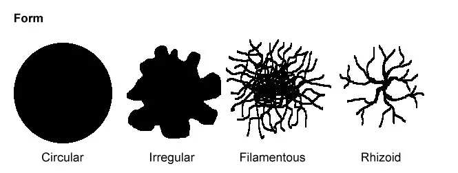
- Shapes that are irregular, rhizoid, filamentous, or spherical.
- Colonies can be elevated, flat, convex, umbonate, pulvinate, or crateriform depending on their elevation.
- Edges might be whole, lobate, crenate, undulate, or ciliate.
- Color – Specific bacterial species create pigments. Serratia marcescens produces the orange-red pigment known as prodigiosin. Pseudomonas aeruginosa produces the pigments pyoverdin and pyocyanin, which give the colonies a greenish sheen.
- Transparent, translucent, or opaque describes opacity.
- Haemolysin, a substance made by some bacteria, causes hemolysis close to the colony. Accordingly, the bacterial colony could be:
- Alpha – Blood cells are partially destroyed.
- Blood cells are destroyed in beta.
- Gamma – Blood cells are not destroyed.
Colony shapes
It includes the bacterial colony’s shape, elevation, and margin.
The shape of the bacterial colony is referred to as its form. These four types are the most typical colony shapes you will probably see.
- Rhizoid
- Filamentous
- Circular
- Irregular

Elevation of the bacterial colony: It reveals the colony’s height above the agar. This is how a colony appears from the “side.”
There are six main bacterial colony elevations:
- Crateriform
- Convex
- Flat
- Elevated
- Umbonate (with a knobby protuberance)
- Pulvinate (cushion-shaped).

The edge or margin of a bacterial colony may be a key element in determining an organism’s identity. Examples of this
- Lobate
- Curled
- Whole (smooth)
- Uneven
- Undulate (wavy)
- Filiform.
Colonies with uneven shapes and/or edges are probably made up of mobile creatures. Proteus spp. and other highly mobile species inundated the culture media.

Size of the bacterial colony
The colony’s size may be a helpful characteristic for identification. A representative colony’s diameter can be expressed in relative terms like pinpoint, small, medium, and big or in millimeters.
Bacterial colonies that are punctiform and various shapes
Punctate colonies are another name for small colonies (pin-point). Colonies that are greater than 5 mm are probably made up of moving creatures. Punctiform colonies are exceedingly tiny, which sets them apart from circular colonies.
The surface of the colony’s appearance
Colonies of bacteria usually have a shiny, smooth look. Other surface adjectives include veined, rough, wrinkled (or shriveled), dull (the reverse of gleaming), and shining. Dry, wrinkly colonies are produced by Bacillus species. Additionally, Pseudomonas stutzeri produces colonies that look wrinkled and similar.
A colony’s color (pigmentation)
Some bacteria produce color as they develop in the medium, such as the green pigment made by Pseudomonas aeruginosa, the buff colonies of Mycobacterium tuberculosis in L.J. media, and the red colonies of Serratia marcescens.
The bacterial colony’s opacity
A bacterial colony’s opacity can be categorized as transparent (clear), opaque (not transparent or clear), translucent (nearly clear but vision is affected, like looking through frosted glass), or iridescent (changing colors in reflected light). On blood agar, tiny, translucent colonies that are -hemolytic are almost always caused by a Streptococcus species. On the surface of the agar plate, staphylococci produce opaque, smooth, and round colonies.
What purpose does colony morphology serve?
Identification of bacterial species depends heavily on colony shape and its traits. Different bacterial species form colonies of various colors, sizes, shapes, and textures. It is also the accepted approach for identifying the bacterium based solely on appearance.
Describe hemolysis
Hemolysis, which causes the release of hemoglobin from the RBCs or red blood cells, is the breakdown of the red blood cell membrane by the bacterial protein hemolysin. Hemolytic proteins are found in a variety of bacterial species. i.e; Streptococcus pneumonia.
References:
https://byjus.com/neet/colony-characteristics-of-bacteria/
https://microbeonline.com/colony-morphology-bacteria-describe-bacterial-colonies/


