History of digital microscope
1986 saw the creation of an early digital microscope by the now-known Hirox Co. LTD company, which was based in Tokyo, Japan. It had a lens attached to a computer and a control box. Initially, an analogue S-video connection was used to connect to the computer. The connection was eventually upgraded to FireWire 800 in order to manage the substantial amount of digital data arriving from the digital camera.
Advanced all-in-one systems with a monitor and computer built in were first developed in 2005, eliminating the need for a separate computer. Later, in late 2015, they unveiled a system that, while still having the computer separate, connected to it through USB 3.0 to take advantage of its speed and robustness.
This system’s physical size and quantity of wires were both significantly smaller than those of earlier iterations. Some pupils in Laos can view insect parts with a digital microscope. This model was around $150 USD.
The development of the USB port led to the creation of numerous USB microscopes with varying levels of quality and magnification. They keep getting cheaper, especially when compared to conventional optical microscopes. They provide high-resolution images that are typically directly recorded to a computer and that also use the computer’s power for an LED light source that is incorporated into the device. The number of megapixels that are available on a particular model, ranging from 1.3 MP to 2 MP to 5 MP and above, directly affects the resolution.
What is a digital microscope?
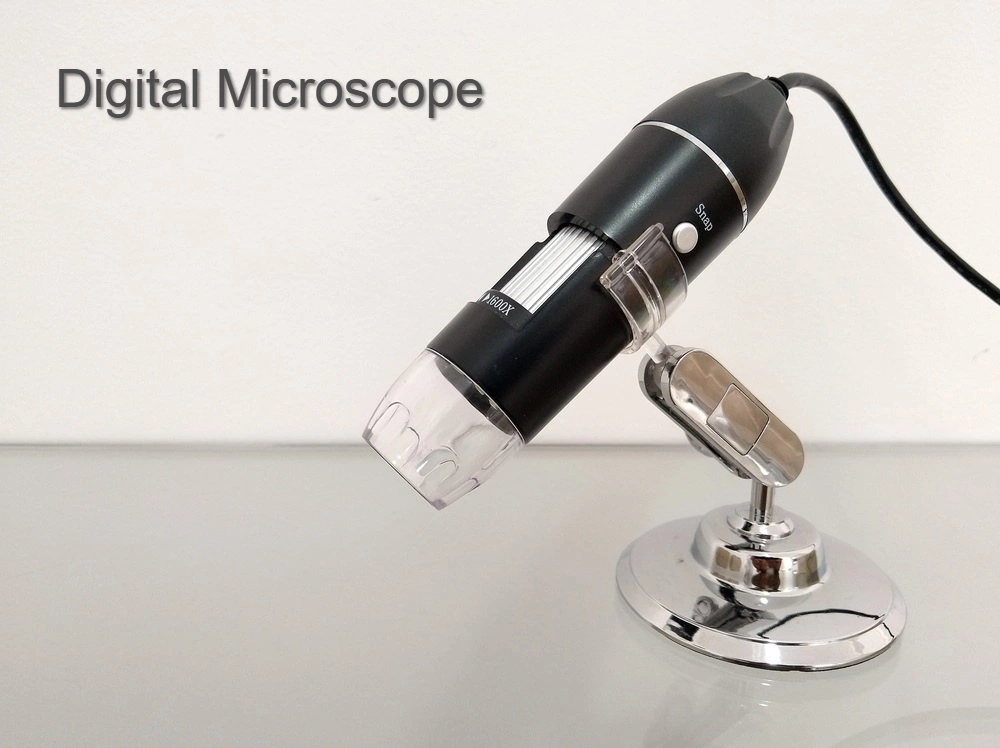
A digital microscope is a modification of a conventional optical microscope that outputs an image to a monitor using optics and a digital camera, occasionally using computer software. Unlike an optical microscope, which allows for direct observation of the sample through an eyepiece, a digital microscope frequently has its own built-in LED light source. The entire system is built for the monitor image since the image is concentrated on the digital circuit. The human eye’s optics are not included.
Digital microscopes range in price from basic USB models that are often affordable to high-end industrial models that cost tens of thousands of dollars. Low cost commercial microscopes typically lack illumination optics (such as Köhler illumination and phase contrast lighting) and resemble webcams with macro lenses more. A digital camera can be added to an optical microscope.
What are the characteristics of digital microscope?
A digital camera’s ability to magnify an image depends on the size of the monitor it is displayed on, making it more powerful than the typical optical microscope. Multiplying the eyepiece and objective lens magnifications yields the power of magnification in a conventional microscope.
Users in every field who spend hours a day working with a microscope benefit from the ergonomics of using a digital microscope. There is no longer any need for viewers to look through an eyepiece, allowing them to sit comfortably upright. Digital microscopes can be connected to a computer monitor and then projected for audience or conference viewing through an LCD projector onto a huge screen.

What are the parts of digital microscope?
There are some fundamental components of a digital microscope, despite the fact that there are undoubtedly many distinct varieties and applications for them. Digital microscope cameras are available in some optical microscopes, and some optical microscopes can output through HDMI or USB. While many other digital microscopes rely on a separate USB computer monitor or HDMI monitor, several digital microscopes incorporate a digital microscope camera with an LCD screen adapter for presenting images.
Types of digital microscope
Handheld microscopes are one of the various varieties of digital microscopes that are available for use in educational, medical, and industrial research. They are perfect for usage in the field and on manufacturing floors in a variety of sectors since they are light and simple to hold.
There are many different industries and associated applications for which digital microscopes have been specially created. Dentiscopes and ear scopes are two examples of digital devices designed for particular uses. Other examples of the numerous varieties of digital microscopes include the following:
High magnification biological microscopes that are lighted from beneath the mechanically graduated stage are known as biological digital microscopes. Using halogen or LED illumination, objectives vary from 4x to 100x.
Fluorescence Digital Microscopes:
Optical instruments produce images primarily from the light emitted by fluorescence and phosphorescence.
Inverted digital microscopes:
Inverted digital microscopes are available in metallurgical, brightfield, and phase contrast formats. These are trinocular microscopes with the light source and condenser on top of the stage.
Metallurgical digital microscopes
These instruments are used to examine circuitry, metal surfaces, and other opaque surfaces.
Phase Digital Microscopes
These upright or inverted microscopes are used to view living or dead specimens that are not stained.
Digital polarising microscopes
These microscopes use polarised light to direct the vibration of light waves in a certain direction. This enables you to assess the anisotropic specimen’s three-dimensional structure and makeup.
Stereo digital microscopes:
Usually, instead of shining light through the specimen, they reflect it back. Excellent for examining plants, circuitry, electrical components, works of art, and antiquities.
Microscopes with USB connections:
Digital microscopes have a separate camera for the microscope. The digital camera is fixed in place and cannot be taken off. Digital microscope packages are cameras that can be disconnected from the microscope to which they are attached using a C-mount adapter.
Handheld digital microscope:
Digital cameras that are newly integrated into compact, handheld microscope systems are known as handheld digital microscopes. utilised in fields like forensics and surface inspection.
Portable digital microscopes:
Conveniently small and frequently equipped with wireless capabilities, portal digital microscopes are frequently used in dermatology, field inspection, and healthcare to observe challenging surfaces.
Digital eyepiece for microscopes:
A variety of optional accessories included in the software serve multiple purposes, including phase contrast observation, bright and dark field observation, microphotography, image processing, particle size determination in m, pathological report and patient manager, microphotography, recording mobility video, drawing and labeling etc.
Resolution:
Typically, a 1600 by 1200 pixel image is produced using a 2 megapixel CCD. The field of view of the camera’s lens affects the resolution of the image. The horizontal field of vision (FOV) can be calculated by dividing it by 1600 to get an approximate idea of the pixel resolution.
A sub-pixel image can be produced to increase resolution. The CCD is physically moved by an actuator in the Pixel Shift Technique in order to capture numerous overlapping images. Sub-pixel resolution can be produced in the microscope by merging the images. This technique gives sub-pixel information; another effective technique is to average a reference image.
2D measurement:
The majority of high-end digital microscope systems can measure samples in two dimensions. Onscreen measurements are made by counting the distance between each pixel. This permits measurements of length, width, diagonal, and circles, among many other things. Some systems can even count the number of particles.
3D measurement:
By stacking images, a digital microscope can measure objects in three dimensions. The system captures images from the lowest focal plane in the field of view to the highest focal plane using a step motor. Finally, a 3D colour image of the sample is created by reconstructing these photos into a 3D model based on contrast. It is possible to take measurements from this 3D model, however the precision depends on the step motor and the depth of field of the lens.
2D and 3D tilling:
The more sophisticated digital microscope systems can now perform 2D and 3D tiling, often known as stitching or making a panoramic image. By using 2D tiling, the XY stage is moved to automatically and flawlessly tile the image. To create a 3D panorama, 3D tiling combines the Z-axis movement of 3D measuring with the XY stage movement of 2D tiling.
What are the advantages of digital microscope?
The instrument’s ergonomics are undoubtedly its most noticeable benefit. Users can view the sample image immediately and use software to evaluate it while seated comfortably and erect because the sample image is presented on a monitor. When dealing with high sample throughput or when users spend a lot of time each day using microscopes, the ergonomics of digital microscopes are very helpful.
Some digital microscopes also come with software that supports the saving of numerous user profiles.This feature is extremely helpful if several people use the same microscope because it allows each person to start working right away with little to no alterations to the microscope working station by simply selecting their own microscope profile.
What are the limitations of digital microscope?
When opposed to stereo or compound microscopes, for example, a digital microscope’s obvious drawback is the requirement for a power connection. For digital microscopes, the picture of the sample is always shown on a monitor because there are no eyepieces. Hence, a minimum of one power cable is required. Digital microscopes typically also require a PC connection or, at the very least, a built-in viewing screen. Customers still have the choice to view their sample through eyepieces when using traditional microscopes.
References
- https://www.leica-microsystems.com/science-lab/applied
- https://microscopeinternational.com/what-is-a-digital-microscope/
- https://tagarno.com/what-is-a-digital-microscope/
- https://en.wikipedia.org/wiki/Digital_microscope


